The cell cycle is the series of events that takes place in a cell, leading to its division and duplication (replication). In cells without a nucleus ( prokaryotic cells e.g. bacteria), the cell cycle occurs through a process termed binary fission. In cells with a nucleus (eukaryotes ), the cell cycle can be divided in two brief periods: interphase —during which the cell grows, accumulating nutrients needed for mitosis and duplicating its DNA —and the mitosis phase, just after interphase, in which the cell splits itself into two distinct cells, often called "daughter cells". The cell-division cycle is a vital process by which a single-celled fertilized egg develops into a mature organism, as well as the process by which hair, skin, blood cells, some internal organs are renewed and wounds are healed.
MITOSIS:
Interphase (pre-mitosis): The cell spends most of its life in the interphase. During this phase the cell grows to its maximum size and performs its normal functions. Many scientists do not count interphase as part of mitosis.
 Prophase: The DNA condenses into chromosomes (human cells have 46 chromosomes – 23 from your father and 23 from your mother). Each chromosome eventually can be seen to consist of two strands or chromatids joined at a central centromere in an X shape. The nuclear membrane disappears. An organelle called the centriole splits and starts to move to opposite poles. Spindle threads form between the poles.
Metaphase: Chromosomes lie on the equator of the cell. Each chromosome is attached to the spindle microfibers by its center. The chromosomes appear in a straight line across the middle of the cell.
Anaphase: The center of the chromosome splits. Each chromosome divides into two sister chromatids. Each chromatid is moved to opposite poles of the cell by the shortening of the spindle fibres. Chromatids (now called daughter chromosomes ) gather at opposite poles of the cell.
Telophase: A nuclear membrane forms around each of the daughter chromosomes that have gathered at the poles. The daughter chromosomes uncoil. The cytoplasm then divides during a process called cytokinesis . Note –cytokinesis is not a stage of mitosis but the process of the cytoplasm splitting into two. There are now two genetically identical daughter cells. They are identical to the parent cell and to each other.
Biological importance of mitosis
- Growth – Living tissue grows by mitosis e.g. bone and skin.
- Repair - Damaged and worn-out tissues are replaced with new cells by mitosis.
- Asexual reproduction - Single-celled (unicellular) organisms and bacteria often reproduce asexually by mitosis. Organisms like amoeba are able to split from a single individual into two and therefore can reproduce without a mate and sexual reproduction.

This text is adapted under a Creative Commons 4.0 license. You can find the original source here.
More information can be found on the powerpoint.
Molecular Genealogy - Tracing DNA through a Family Tree
 As prophase I progresses, the close association between homologous chromosomes begins to break down, and the chromosomes continue to condense, although the homologous chromosomes remain attached to each other at chiasmata (crossing over point). At the end of prophase I, the pairs are held together only at chiasmata and are called tetrads because the four sister chromatids of each pair of homologous chromosomes are now visible.

Step Thru Animation

Molecular Genealogy - Tracing DNA through a Family Tree
MEIOSIS:
Introduction
Sexual reproduction requires fertilization, a union of two cells from two individual organisms. If those two cells each contain one set of chromosomes, thus the resulting cell contains two sets of chromosomes. Haploid cells contain one set of chromosomes. Cells containing two sets of chromosomes are called diploid. If the reproductive cycle is to continue, the diploid cell must somehow reduce its number of chromosome sets before fertilization can occur again, or there will be a continual doubling in the number of chromosome sets in every generation. So, in addition to fertilization, sexual reproduction includes a nuclear division, known as meiosis, that reduces the number of chromosome sets.
The nuclear division that forms haploid cells, which is called meiosis, is related to mitosis. As you have learned, mitosis is part of a cell reproduction cycle that results in identical daughter nuclei that are also genetically identical to the original parent nucleus. In mitosis, both the parent and the daughter nuclei contain the same number of chromosome sets—diploid for most plants and animals. Meiosis employs many of the same mechanisms as mitosis. However, the starting nucleus is always diploid and the nuclei that result at the end of a meiotic cell division are haploid.
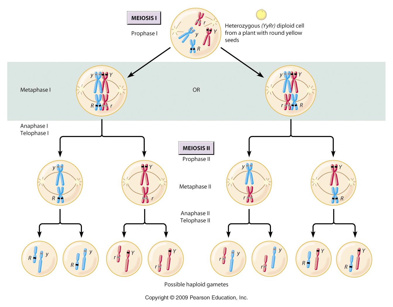
In order to cut the chromosome number in half, meiosis consists of one round of chromosome duplication and two rounds of nuclear division. The first round on division is called meiosis I, whereas the second division is called meiosis II. The genetic information is mixed during the first division to create unique recombinant chromosomes.
Meiosis I
As the nuclear envelope begins to break down, the homologous chromosomes line up. An exchange of chromosome segments between non-sister homologous chromatids occurs and is called crossing over.
The crossover events are the first source of genetic variation produced by meiosis. A single crossover event between homologous non-sister chromatids leads to a reciprocal exchange of equivalent DNA between a maternal chromosome and a paternal chromosome. Now, when that sister chromatid is moved into a gamete, it will carry some DNA from one parent of the individual and some DNA from the other parent. The recombinant sister chromatid has a combination of maternal and paternal genes that did not exist before the crossover.
During metaphase I, the homologous chromosomes are arranged in the center of the cell. The orientation of each pair of homologous chromosomes at the center of the cell is random.


During metaphase I, the homologous chromosomes are arranged in the center of the cell. The orientation of each pair of homologous chromosomes at the center of the cell is random.
This randomness, called independent assortment, is the physical basis for the generation of the second form of genetic variation in offspring. Consider that the homologous chromosomes of a sexually reproducing organism are originally inherited as two separate sets, one from each parent. Humans have 23 chromosome pairs, which results in over eight million possible arrangements of the chromosomes in the daughter cells. This number does not include the variability previously created in the sister chromatids by crossover. Given these two mechanisms, it is highly unlikely that any two haploid cells resulting from meiosis will have the same genetic composition.
To summarize the genetic consequences of meiosis I: the maternal and paternal genes are recombined by crossover events occurring on each homologous pair during prophase I; in addition, the random assortment of tetrads at metaphase produces a unique combination of maternal and paternal chromosomes that will make their way into the gametes.
In anaphase I, the spindle fibers pull the linked chromosomes apart. The sister chromatids remain tightly bound together at the centromere. It is the chiasma connections that are broken in anaphase I as the fibers attached to the fused kinetochores pull the homologous chromosomes apart.
In telophase I, the separated chromosomes arrive at opposite poles.
Cytokinesis, the physical separation of the cytoplasmic components into two daughter cells, occurs. At each pole, there is just one member of each pair of the homologous chromosomes, so only one full set of the chromosomes is present. This is why the cells are considered haploid—there is only one chromosome set, even though there are duplicate copies of the set because each homolog still consists of two sister chromatids that are still attached to each other. However, although the sister chromatids were once duplicates of the same chromosome, they are no longer identical at this stage because of crossovers.
Meiosis II
In meiosis II, the connected sister chromatids remaining in the haploid cells from meiosis I will be split to form four haploid cells. Chromosomes are not duplicated during meiosis II. The two cells produced in meiosis I go through the events of meiosis II in synchrony. Overall, meiosis II resembles the mitotic division of a haploid cell.
In prophase II, the centrosomes move away from each other toward opposite poles, and new spindles are formed. Each sister chromatid forms an individual kinetochore that attaches to microtubules from opposite poles. In metaphase II, the sister chromatids are maximally condensed and aligned at the center of the cell. In anaphase II, the sister chromatids are pulled apart by the spindle fibers and move toward opposite poles.
In telophase II, the chromosomes arrive at opposite poles and begin to decondense. Nuclear envelopes form around the chromosomes. Cytokinesis separates the two cells into four genetically unique haploid cells. At this point, the nuclei in the newly produced cells are both haploid and have only one copy of the single set of chromosomes. The cells produced are genetically unique because of the random assortment of paternal and maternal homologs and because of the recombination of maternal and paternal segments of chromosomes—with their sets of genes—that occurs during crossover.
Step Thru Animation
Comparing Meiosis and Mitosis
Mitosis and meiosis, which are both forms of division of the nucleus in eukaryotic cells, share some similarities, but also exhibit distinct differences that lead to their very different outcomes.
Mitosis is a single nuclear division that results in two nuclei, usually partitioned into two new cells. The nuclei resulting from a mitotic division are genetically identical to the original. They have the same number of sets of chromosomes: one in the case of haploid cells, and two in the case of diploid cells.
On the other hand, meiosis is two nuclear divisions that result in four nuclei, usually partitioned into four new cells. The nuclei resulting from meiosis are never genetically identical, and they contain one chromosome set only—this is half the number of the original cell, which was diploid.
The differences in the outcomes of meiosis and mitosis occur because of differences in the behavior of the chromosomes during each process. Most of these differences in the processes occur in meiosis I, which is a very different nuclear division than mitosis. In meiosis I, the homologous chromosome pairs become associated with each other, are bound together, experience chiasmata and crossover between sister chromatids, and line up along the metaphase plate in tetrads with spindle fibers from opposite spindle poles attached to each kinetochore of a homolog in a tetrad. All of these events occur only in meiosis I, never in mitosis.
Homologous chromosomes move to opposite poles during meiosis I so the number of sets of chromosomes in each nucleus-to-be is reduced from two to one. For this reason, meiosis I is referred to as a reduction division. There is no such reduction in ploidy level in mitosis.
Meiosis II is much more analogous to a mitotic division. In this case, duplicated chromosomes (only one set of them) line up at the center of the cell with divided kinetochores attached to spindle fibers from opposite poles. During anaphase II, as in mitotic anaphase, the kinetochores divide and one sister chromatid is pulled to one pole and the other sister chromatid is pulled to the other pole. If it were not for the fact that there had been crossovers, the two products of each meiosis II division would be identical as in mitosis; instead, they are different because there has always been at least one crossover per chromosome. Meiosis II is not a reduction division because, although there are fewer copies of the genome in the resulting cells, there is still one set of chromosomes, as there was at the end of meiosis I.
Cells produced by mitosis will function in different parts of the body as a part of growth or replacing dead or damaged cells. They may even be involved in asexual reproduction in some organisms. Cells produced by meiosis in a diploid-dominant organism such as an animal will only participate in sexual reproduction.
Add a comment